
Nanolithography-Enabled Sensor Arrays for Rapid Cancer Detection
-
Author:
Hui-Yu Liu
-
Source:
Dissertation, Westfälische Wilhelms-Universität (WWU) (2020)
- Date: 2020
-
Cancer is the second leading cause of death globally after cardiovascular diseases, accounting for an estimated 9.6 million deaths in 2018 (WHO). Cancer can start in almost any organ or tissue of the body when abnormal cells grow uncontrollably, go beyond their usual boundaries to invade other parts of the body and spread to other organs via the circulatory systems. The latter process is commonly referred to as metastasis and as it progresses it both greatly increases the difficulty of fighting the cancer and forms a major cause of death. An investigation of the WHO shows that the one-year survival rate decreased with the increasing stages of progression of the cancer at diagnosis. Major reductions were observed for stage 4 breast-, prostate- and colorectal cancer. It is therefore of utmost importance for the chances of survival that cancer is diagnosed during early stages of development. Cancer is traditionally diagnosed through a combination of scanning techniques (such as CT and MRI) and invasive biopsy. This method requires the tumor to be detectable by the scanning techniques, is expensive, time-consuming, and causes patient uncomforted. Lab-on-a-Chip technologies have been developed for over three decades as a possibly alternative to detect cancer by a non-invasive liquid biopsy. This technology has the potential of becoming a fast, affordable and accessible method for early cancer detection, but also to help develop personalized treatment plans for patients. This thesis presents research into improving lab-on-a-chip devices by optimizing the microarray and structure of the micro-fluidic chips and by investigating novel methods to detect alternative indicators of cancer. The research has thereby led to new tools for early diagnosis of cancer and establishes a platform for the detection of various other disease, such as Alzheimer’s disease and cardiovascular diseases.
Chapter 1.1 introduces the evolution of nanolithography that is applied in the scope of this work. In brief, dip pen nanolithography (DPN) was first introduced about two decades ago and has since then gradually developed into several DPN-based technologies, among which polymer pen lithography (PPL) and DPN with lipid inks (L-DPN). Phospholipids are the main compounds in cell membranes. Mimicking phospholipid bilayers is therefore very enabling in biology research. DPN can easily generate these supporting phospholipid bilayers on various kinds of substrates. The technique of polymer-pen lithography (PPL) belongs to the family of scanning probe lithography techniques. It is an inexpensive, cantilever-free technique with micro- to nanoscale resolution. It is a hybrid technique of DPN and microcontact printing (μCP). PPL combines the key capabilities of both techniques, i.e. large area patterning astypically achieved in μCP and the precise control of features such as shape and size from DPN. The techniques of PPL and L-DPN were applied in fabricating microarray biosensors for cancer in my research.
The previous study [1] established the circulating tumor cell (CTC) microfluidic chips which is a novel system of CTC capture by microarray surfaces. Sensitized tumor cells from a culture are spiked into a blood sample of a healthy donor. A custom-made microfluidic chip ensures that the cells come sufficiently into contact with the microstructure located at the bottom of the device by a staggered herringbone (SHB) structure. Only sensitized cells are immobilized on the microarrays through the process of antibody binding. Healthy blood cells pass by the chip. The result showed the approach recovered almost 50% of the tumor cells at a specificity of 85 %. In chapter 2.1, in order to optimize the system (CTC microfluidic chips) for future clinical use, different patterns of PPL and staggered herringbone (SHB) designs were tested. The Parsortix system of enrichment equipment which uses the size differences of cells to separate CTCs and blood cells in blood samples, was combined with the chips for spiked samples. The optimized system (HC chips) shows nearly full capture rate and 90% viability of captured cells, which indicates the optimized system could not be used just for detecting cancer, but also for CTCs isolation and downstream analysis.
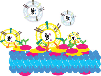
EVs are generated by cells, they carry abundant information (DNA, RNA, proteins) for cell communications. Cancer cells are proven to produce much more EVs than healthy cells. In cancer progress and metastasis, cancer cells use EVs to affect healthy cells, for instance, to affect immune cells to not recognize cancer cells, and to control healthy bone marrow cells to build the new niche in the target organ for preparing metastasis. Hence, cancer EVs are key in cancer progress. Furthermore, cancer EVs are shown to carry specific cancer markers, which can be used as biomarkers in liquid biopsy for cancer detection. However, before cancer EVs can be routinely used in clinical and liquid biopsy analysis, reliable and highly sensitive methods with high specificity must be developed for their isolation from and detection in complex backgrounds (such as blood). In chapter 2.2, we use L-DPN to establish a unique method for capturing cancer EVs. The lipid arrays are functionalized by cancer antibodies for specific capturing. I use the special lipidic property of fusion between lipid arrays and cancer EVs, and the immunoaffinity of antibodies to trap the targeted cancer EVs. The results show cancer EVs can be caught by our lipid arrays. Furthermore, the EV-derived RNA has been proven to be trapped in our lipid patches, which can help downstream analysis for cancer research. Our EV-lipid array therefore is not only a device for detection, but also a device for isolation.
This thesis investigates the development of microarrays in chips for cancer detection by polymer pen lithography (PPL) and dip-pen nanolithography (DPN). The results show that our CTC chip and EV-lipid array are efficient tools for clinical cancer detection. Moreover, the applicability of our devices is not limited to cancer detection. The CTC chip and EV-lipid array are platforms for detection and isolation of rare cells and extremely small EVs in a complex background, which can also apply in detection of different diseases.