
4D-STEM
4D-STEM techniques enable correlated and simultaneous analysis of structural and functional characteristics at the nanoscale combining pair distribution function or crystal orientation analysis with measuring strain and magnetic or electric fields.
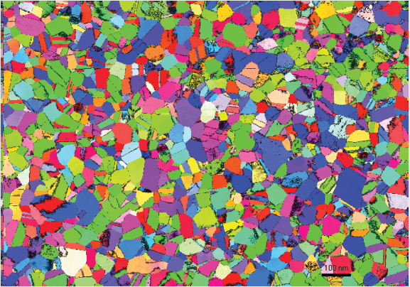
Quantitative Automated Crystal Orientation Mapping in STEM mode
link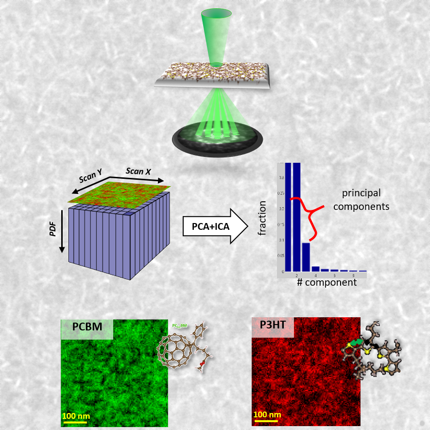
Pair distribution function mapping in STEM to image the phase distribution and structure of amorphous nanocomposites.
link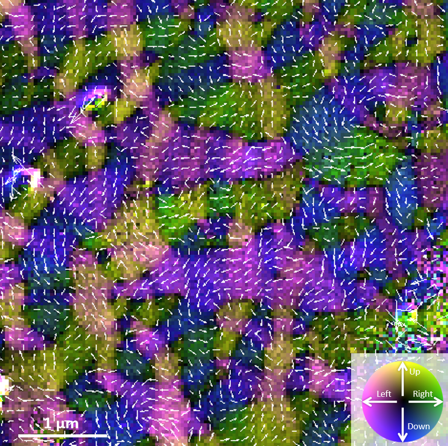
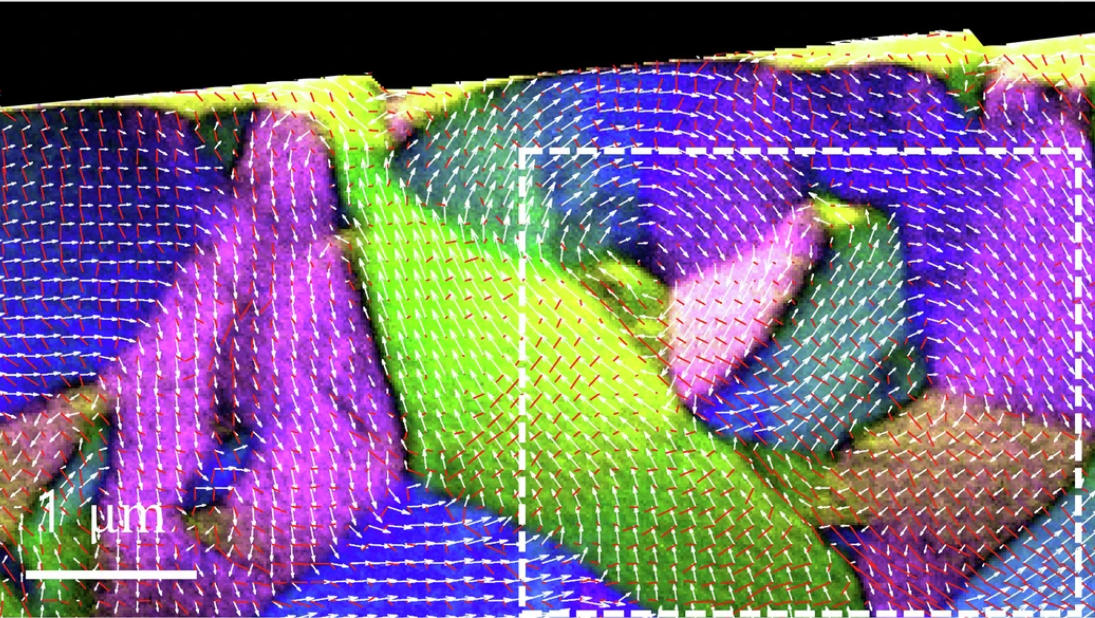
Differential phase contrast imaging in STEM mode is one of the most powerful methods to image magnetic fields.
linkTecnique
4D-STEM is combining scanning in STEM mode with the acquisition of full nanobeam diffraction patterns at every scan position, creating a 2D array of 2D diffraction patterns. The diffraction patterns contain both structural [1,2,3,4] and functional [5] information, which can be analyzed to retrieve both simultaneously from the same data set. This enables the most direct correlation of structural and functional properties and can further be readily combined with in-situ approaches to evaluate their dynamic evolution [1,6,7].
For crystalline materials, ACOM-STEM is well established yielding phase and orientation maps of materials with nanometer resolution [1,6,7]. This can be extended by Cepstral analysis for high quality strain mapping. The counterpart for amorphous materials is STEM-PDF [2,4,5], where the local pair distribution function provides information on short and medium ranger order in metallic glasses, disordered ceramics and organic materials. Analysis of the ellipticity of the diffraction halos further enables local strain analysis in these glassy materials [4,5].
The displacement of the diffraction patterns provides information on local electric and magnetic fields inside the sample. Using a singular valued decomposition approach [4] provides extreme sensitivity for measuring these displacements and thus access to small electric or magnetic fields [5]. Moreover, this approach suppresses most of the crystallographic artifacts enabling a precise measurement of the local fields. Because of the high sensitivity, it can be used to analyze typical ACOM-STEM and STEM-PDF data sets, thus giving access to simultaneous and correlated structural and functional information.
_____________________________________________________________________
Details and further work are published at:
-
Combination of in-situ straining and ACOM TEM: a novel method for analysis of plastic deformation of nanocrystalline metals
A. Kobler, A. Kashiwar, H. Hahn, C. Kübel
Ultramicroscopy, 2013, 128, 68-81; DOI: 10.1016/j.ultramic.2012.12.019. -
Radial distribution function imaging by STEM diffraction: Phase mapping and analysis of heterogeneous nanostructured glasses
X. Mu, D. Wang, F. Tao, C. Kübel
Ultramicroscopy, 2016, 168, 1-6; DOI: 10.1016/j.ultramic.2016.05.009. -
Mapping structure and morphology of amorphous organic thin films by STEM pair distribution function analysis
X. Mu, A.A. Mazilkin, C. Sprau, A. Colsmann, C. Kübel
Microscopy, 2019, 68(4), 301-309; DOI: 10.1093/jmicro/dfz015. -
Direct observation of quadrupolar strain fields surrounding Eshelby inclusions in metallic glasses
S.J. Kang, D. Wang, A. Caron, C. Minnert, K. Durst, C. Kübel, X. Mu
Advanced Materials, 2023, 35, 2212086; DOI: 10.1002/adma.202212086. -
Large-angle Lorentz 4-Dimensional Scanning Transmission Electron Microscopy for Simultaneous Local Magnetic, Strain and Structural Mapping
S. Kang, M. Töllner, D. Wang, C. Minnert, K. Durst, A. Caron, R.E. Dunin-Borkowski, J. McCord, C. Kübel, X. Mu
Nature Communication, 2025, 16, 1305; DOI: 10.1038/s41467-025-56521-6. -
In situ observation of deformation processes in nanocrystalline face-centered cubic metals
A. Kobler, C. Brandel, H. Hahn, C. Kübel
Beilstein Journal of Nanotechnology, 2016, 7, 572–580; DOI: 10.3762/bjnano.7.50. -
In situ TEM observation of cooperative grain rotations and the Bauschinger effect in nanocrystalline palladium
A. Kashiwar, H. Hahn, C. Kübel
Nanomaterials, 2021, 11, 432; DOI: 10.3390/nano11020432.