Correlative Characterization

Our research focuses on correlative characterization workflows that integrate complementary scale bridging imaging techniques such as FIB/SEM, light microscopy, X-ray CT, and TEM into a unified 3D representation of the material.

Dr. Christian kübel

Correlative characterization is essential for scale bridging analysis and to probe different properties and features.

We are developing approaches for 3D correlative characterization enabling a transfer between different instruments from optical microscopy to X-ray CT, FIB, TEM and APT to address different length scales and to combine surface analysis such as AFM, XPS and ToF-SIMS with electron microscopy techniques.

In addition, we are using and developing solutions for in-instrument correlation of different techniques.

  • 3D/4D STEM correlating PDF analysis, DPC magnetic domain imaging and strain mapping in addition to the well established imaging with EELS and EDX spectroscopy.

  • Nanovision AFM-in-SEM combining AFM topology analysis with SEM/FIB, nanoindentation and electrical measurements.

Correlative Characterization Across Length Scales

Correlated analysis by 4D-STEM Sangjun Kang and Xiaoke Mu
4D-STEM

4D-STEM based local correlated analysis of pair distribution function (PDF), differential phase contrast (DPC) and strain.

NenoVision Litescope NenoVision
AFM-in-SEM

Nenovision LiteScope: AFM (topography, tapping mode), nanoindentation, C-AFM, KPFM, STM, MFM, PFM

link

Correlative 3D Workflow


Figure 2- Our correlative workflow allows targeting and extracting a sub-surface region of interest (ROI, several µm) within a macroscopic sample (mm to cm), with an accuracy of 1-2 µm - essentially a needle-in-a-haystack problem. Global markers, local markers and ROI markers are used for tracking and registering the ROI in 3D.

Correlative Microscopy


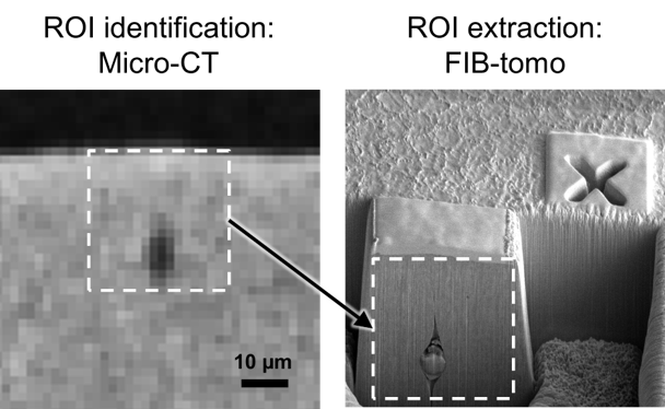
A central challenge in correlative microscopy is the accurate localization and revisiting of identical regions of interest across modalities with fundamentally different imaging geometries and contrasts.
To solve this problem, we developed a two-step alignment and ROI targeting concept based on global, local, and fine reference markers, supported by a dedicated sample carrier. This enables robust three-dimensional registration and micrometer-scale targeting accuracy, including for sub-surface and weak-contrast features. We apply these methods to address challenging materials science questions and collaborate with partners to implement them in diverse experimental environments.


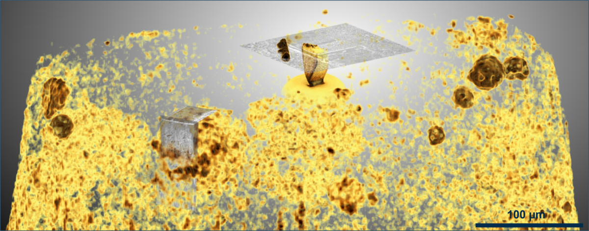
Figure1- FIB tomography (Slice&View) on 30 x 30 x 30 µm² volume, showing an encapsulated particle in extruded steel, initially identified by X-ray Micro-CT. See the Focused Ion Beam page.