Porous Materials
Visualizing Confinement: Quantification of Transport in Porous Materials
Porous materials are often described in terms of surface area and average pore size. Yet at the nanometer scale, where mesoporous silicas operate, molecular transport is governed less by averages than by the precise three-dimensional architecture of the pore network. Constrictions, dead ends, connectivity, and local heterogeneities determine whether diffusion proceeds smoothly or becomes severely hindered. Understanding this interplay requires more than bulk characterization—it requires direct visualization of the pore space itself.
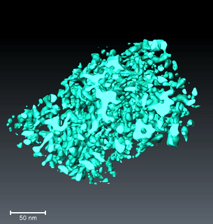
Transmission electron microscopy (TEM), particularly in scanning mode combined with tomographic reconstruction, has emerged as a powerful tool to achieve exactly this. By acquiring tilt series and reconstructing three-dimensional volumes with nanometer resolution, the mesopore space of silica materials can be physically reconstructed and segmented into solid and void phases . These reconstructions are not simply images; they serve as geometrical models for direct pore-scale diffusion simulations.
Unlike classical bulk techniques such as nitrogen physisorption or small-angle X-ray scattering, which require model assumptions to interpret pore structure, tomography provides directly interpretable morphology. From these datasets, quantitative parameters such as void fraction, pore size heterogeneity, connectivity, and the presence of constrictions or closed pores can be extracted. Importantly, statistically significant volumes can be analyzed, enabling morphology to be described across length scales from the nanometer to the sub-micron regime .
To connect structure with function, diffusion of passive finite-size tracers is simulated within the reconstructed pore networks. The key parameter governing transport under confinement is the ratio λ = d_tracer / d_meso, relating solute size to mean pore diameter. As λ increases, steric exclusion reduces the accessible pore volume, and hydrodynamic interactions enhance drag. The effective diffusion coefficient therefore decreases relative to bulk diffusion, a behavior quantified through global and local hindrance factors .
What emerges from this reconstruction–simulation approach is a striking sensitivity of transport to subtle morphological details. In random mesoporous silicas, broad pore size distributions allow larger pores to maintain transport pathways even as smaller pores close off with increasing λ . In ordered materials such as SBA-15 and KIT-6, narrow pore size distributions enhance size selectivity but render diffusion highly susceptible to local imperfections such as constrictions and dead ends . In some cases, diffusion declines sharply at surprisingly small λ values because narrow bottlenecks dominate the network response .
Core–shell particles provide an additional illustration. Designed to decouple diffusion distance from particle size, their mesoporous shells were expected to facilitate transport. However, tomography revealed increased fractions of narrow and closed pores, likely formed during shell consolidation, leading to less favorable connectivity than anticipated . Without three-dimensional reconstruction, such structural penalties would remain hidden behind averaged bulk metrics.
The implications extend directly to catalysis under spatial confinement. In ring-closing metathesis performed within mesoporous silica particles, selectivity depends critically on whether pore diameters permit access of one substrate molecule or allow simultaneous entry of multiple species . Morphology–transport relationships derived from tomography thus provide predictive insight into how pore architecture influences reaction–transport coupling.
By transforming porous materials into digital geometries and solving diffusion directly within them, TEM-based tomography moves characterization beyond descriptive metrics toward predictive understanding. It reveals that transport in mesoporous media is governed not by idealized cylinders but by real, three-dimensional networks whose topology and geometry encode the physics of confinement.
Details and further work are published at:
- D. Stoeckel, C. Kübel, M. Loeh, B. Smarsly, U. Tallarek, Langmuir, 2015, 31(26), 7391–7400; DOI: 10.1021/la5046018.
- Olefin ring-closing metathesis under spatial confinement: Morphology–transport relationships, U. Tallarek, J. Hochstrasser, F. Ziegler, X. Huang, C. Kübel, M.R. Buchmeiser, ChemCatChem, 2021, 13(1), 281-292; DOI: 10.1002/cctc.202001495.
- Morphology–Transport Relationships for SBA-15 and KIT-6 Ordered Mesoporous Silicas, J Hochstrasser, A. Svidrytski, A. Höltzel, T. Priamushko, F. Kleitz, W. Wang, C. Kübel, U. Tallarek, Physical Chemistry Chemical Physics, 2020, 22, 11314; DOI: 10.1039/D0CP01861A.
- Transport under confinement: Hindrance factors for diffusion in core-shell and fully porous particles with different mesopore space morphologies, S.-J. Reich, A. Svidrytski, A. Höltzel, W. Wang, C. Kübel, D. Hlushkou, U. Tallarek, Microporous & Mesoporous Materials, 2019, 282, 188–196; DOI: 10.1016/j.micromeso.2019.02.036.