

Ptychography and Phase Reconstruction
Recovering the phase of the electron wave is essential for imaging light and heavy atoms, local potentials and beam-sensitive materials at high resolution and low dose at the same time. Because electron detectors record only intensities, the phase must be reconstructed computationally. This is a hard inverse problem, especially at low dose and for thicker specimens where multiple scattering breaks simple imaging models. Our group develops, benchmarks and applies phase-reconstruction algorithms for 4D-STEM, recovering the complex exit wave and projected potential from a full diffraction pattern at every probe position.
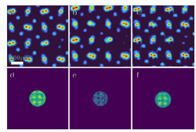
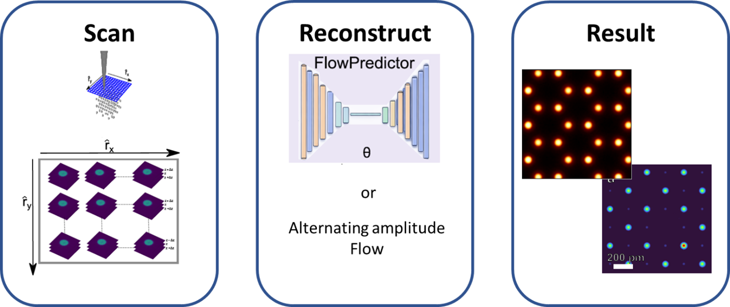
Figure 1. Overview: from a scanned 4D-STEM acquisition to a quantitative projected-phase map of the specimen.
How phase reconstruction works
A pixelated detector stores the entire diffraction pattern at each probe position, so reconstruction is done after acquisition. Each pattern holds only the squared absolute of the exit wave. The phase, where the object’s potential is encoded, must be calculated. Overlapping patterns from neighbouring positions or a defocus series supply the redundancy that makes this inversion possible. We pursue two complementary routes:
a robust iterative ptychography method and a physics-informed machine-learning approach..
- The recorded signal is intensity only, phase must be reconstructed to reveal the projected potential.
- Probe overlap or defocus series supplies the redundancy that makes phase retrieval well-posed.
- Direct methods (SSB, WDD) are fast but tied to the weak-phase-object approximation, iterative and learned methods relax it.
Robust single-slice ptychography with Alternating Amplitude Flow
The Alternating Amplitude Flow (AAF) algorithm minimises an amplitude-based data-fidelity term by gradient descent, using predetermined, computable step sizes that remove most manual tuning. We map its reliable operating range on simulated SrTiO3 (and the lower-symmetry PrScO3), varying probe overlap, thickness, convergence angle, detector angle and dose, then validate on an experimental SnS2 dataset including strongly subsampled scans.
Figure 2. PrScO3 reconstructions at electron doses of 106 e−/Å2(a), 103 e−/Å2(b), and a reconstruction at 3mrad(c). (d) shows the infinite dose signal diffraction pattern at a single position (e) Simulated diffraction pattern acquired at 103 e−/Å2 and (f) the reconstructed diffraction
Probe overlap: stable down to ~25% , well below the ~40% typical of comparable engines, enabling faster, lower-dose scans.
- Thickness & angle: atomic positions identifiable to ~10 nm SrTiO3, reliable down to ~6 mrad convergence and ~25–30 mrad detector angle.
- Dose & noise: structure preserved at 10⁴ e⁻/Ų and partly at 10³ e⁻/Ų, outperforming ePIE at low dose.
- Experiment: on SnS2, AAF resolves tin and sulphur columns without tuning and still reconstructs from 1/9 of the scan positions.
Physics-informed machine learning with the Transport-of-Intensity Equation
Our second route revisits the transport-of-intensity equation (TIE), which links axial intensity variations to phase and combines it with a physics-informed neural network. The model estimates a transverse phase-gradient constrained by the continuity form of the TIE, then reconstructs the exit-wave phase. Training couples phase-gradient supervision with a transport-consistency loss and phase regularization, embedding the imaging physics directly into the learned mapping rather than imposing it afterwards.
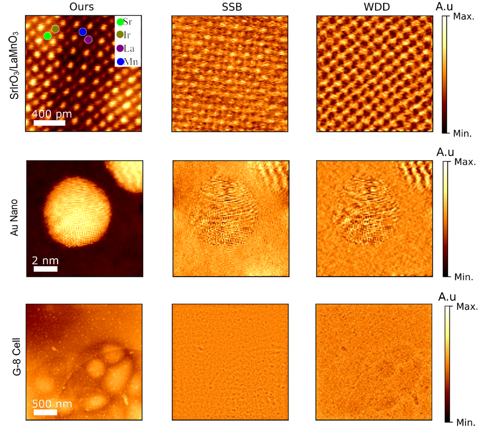
Figure 3. Projected phase reconstruction for experimental data layered SrIrO3/LaMnO3, Gold Nanoparticle, and Cytoplasm of cultured muscle cells using proposed method, SSB, and WDD.
- Physics-constrained learning avoids restrictive weak-scattering or uniform-intensity assumptions.
- Sharper, more localised phase features and improved stability under reduced dose and increasing thickness (SrTiO3, MoS2).
- Generalises to experimental SrIrO3/LaMnO3, Au nanoparticles and biological cells, and serves as a prior for multislice reconstruction.
Details and further work are published at:
- M. Töllner, D. Wang, X. Zhao, H. Scharr, C. Kübel, A. Bangun, “Physics-Based Machine Learning Transport of Intensity Equation for Projected Phase Reconstruction in 4D STEM”, submitted to Small Methods (2026). [Under review]
- M. Töllner, O. Melnyk, X. Mu, D. Wang, T. Lorenzen, K. Müller-Caspary, F. Filbir, C. Kübel, “Robust single-slice electron ptychography using Alternating Amplitude Flow across experimentally relevant parameters”, submitted to Ultramicroscopy (2026). [Under review]
- F. Filbir, O. Melnyk, “Image recovery for blind polychromatic ptychography”, SIAM J. Imaging Sciences 16(3) (2023) 1308–1337. DOI: 10.1137/22M1527155.